
Preventing Oral Surgery Complications in General Dental Practice | WSS
oral surgery complications prevention dental practice

Clinical Education · Western Surgical and Sedation
Surgical complications are a reality of oral surgery practice. They occur in the hands of experienced oral surgeons, and they occur in the hands of well-trained general dentists. The goal of complication management education is not to eliminate the possibility of complications — it is to develop the clinical reflexes that minimize their frequency, recognize them early, and respond to them effectively when they arise.
For general dentists who are expanding their surgical scope or considering doing so, understanding the complication landscape is essential. It informs case selection, shapes the conversations you have with patients during informed consent, and determines whether an unexpected intraoperative finding becomes a managed situation or a crisis.
This article covers the most common complications encountered in third molar surgery and general oral surgical procedures, with practical guidance on prevention, recognition, and management for each.
Dry Socket (Alveolar Osteitis)
Dry socket is the most common postoperative complication following third molar extraction, occurring in approximately 2 to 5 percent of all extractions and up to 30 percent in certain high-risk populations. It results from the premature dissolution or displacement of the blood clot in the extraction socket, exposing underlying bone and nerve tissue.
Clinical presentation
Dry socket presents as increasing pain beginning 2 to 4 days after extraction — characteristically after a period of relative comfort following the procedure. The pain is often described as throbbing and radiating, typically toward the ear or temple. Clinical examination reveals an empty or partially collapsed socket with exposed bone that is visually inspectable and sensitive to air flow.
Risk factors and prevention
Smoking is the most significant modifiable risk factor — nicotine impairs vascular response and clot formation. Patients should be counseled to avoid smoking for a minimum of 48 hours post-extraction, ideally longer.
Oral contraceptive use has been associated with elevated dry socket rates in some populations, likely related to estrogen effects on fibrinolysis.
Traumatic extraction technique — excessive bone removal, prolonged procedure time, aggressive irrigation — increases dry socket incidence. Technique refinement reduces risk.
Poor patient compliance with post-operative instructions — particularly straw use, forceful rinsing, or smoking — is the most common precipitating factor in preventable cases.
Chlorhexidine rinse administered preoperatively has demonstrated modest reduction in dry socket incidence in controlled studies.
Management
Dry socket is managed by gentle irrigation of the socket to remove debris, followed by placement of a medicated dressing — typically a zinc oxide eugenol-based material — directly in the socket. The dressing provides pain relief within minutes in most cases and is changed every 2 to 3 days until the socket begins to epithelialize. Systemic antibiotics are not indicated for uncomplicated dry socket unless signs of infection are present.
Inferior Alveolar Nerve Complications
The inferior alveolar nerve (IAN) runs through the mandibular canal in close proximity to the roots of lower third molars, particularly in mesioangular and horizontal impactions with deep bony positions. IAN complications range from transient paresthesia following routine extraction to persistent hypoesthesia resulting from direct nerve injury.
Radiographic assessment and risk stratification
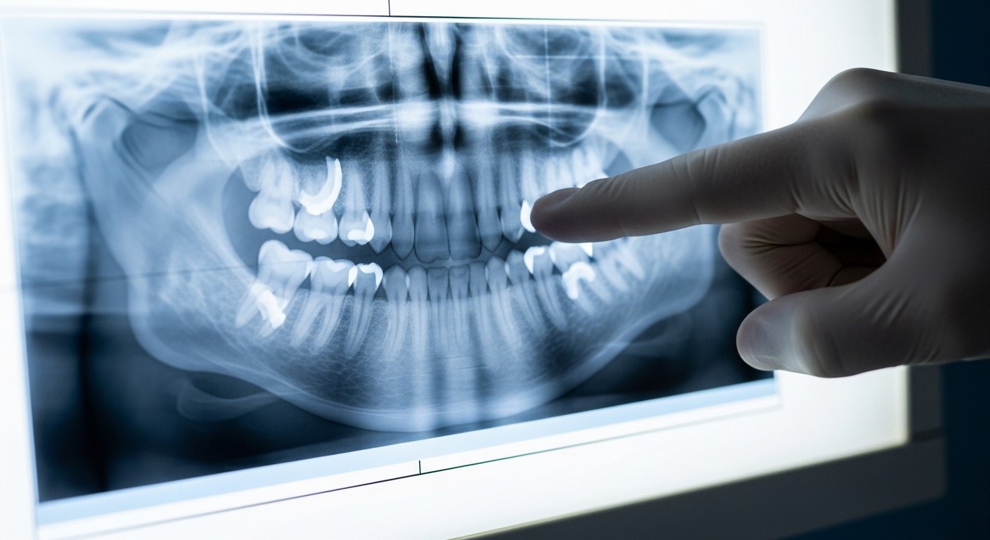
Pre-operative radiographic assessment for IAN proximity is one of the most important risk management steps in lower third molar surgery. Panoramic radiographic signs of high-risk nerve proximity include:
Darkening of the root at the point of canal proximity
Narrowing or deflection of the mandibular canal at the root junction
Interruption of the canal's corticated white line
Grooving or notching of the root surface
When two or more of these signs are present, CBCT imaging should be obtained before proceeding. CBCT provides three-dimensional visualization of the relationship between roots and canal that panoramic radiographs cannot offer, allowing for more accurate risk assessment and better-informed patient consent.
Coronectomy as a risk-reduction technique
In cases where CBCT confirms direct IAN contact and the tooth is otherwise suitable, coronectomy — intentional partial removal that leaves the roots in situ — is an evidence-supported approach that significantly reduces IAN injury risk. The technique involves sectioning the crown from the roots at or below the cemento-enamel junction and leaving the roots to either resorb over time or remain stable. Coronectomy is not appropriate for mobile roots, infected cases, or cases where leaving roots would compromise adjacent structures.
Management of postoperative paresthesia
Transient paresthesia — numbness or tingling in the distribution of the IAN or its branches — following third molar extraction is relatively common and typically resolves within weeks to months. Persistent paresthesia beyond six months warrants specialist referral. Early management includes vitamin B complex supplementation (some evidence supports B6 and B12 in nerve recovery) and patient reassurance with regular follow-up.
Hemorrhage
Postoperative hemorrhage following oral surgery is usually manageable with appropriate pressure and hemostatic technique. Significant hemorrhage that does not respond to local measures is rare but requires a systematic response.
Prevention
Pre-operative screening for bleeding history, anticoagulant use, and platelet disorders reduces the likelihood of hemorrhagic complications. Patients on warfarin, direct oral anticoagulants, or antiplatelet therapy require consultation with their prescribing physician before elective oral surgery. Current evidence supports continuing most anticoagulation regimens for routine oral surgery, as the risk of thromboembolic events from discontinuation generally outweighs the risk of local hemorrhage.
Intraoperative hemostasis
Good surgical technique is the most reliable hemorrhage prevention strategy. Limiting periosteal stripping to what is necessary, managing bone bleeding with bone wax or electrocautery, and closing flaps in tension-free primary closure all reduce postoperative hemorrhage risk. Local hemostatic agents — oxidized cellulose, collagen sponges, or tranexamic acid-soaked gauze — should be available in every surgical kit.
Managing active postoperative hemorrhage
When a patient presents with active postoperative hemorrhage, the first step is direct pressure applied to the site with moistened gauze for a minimum of 20 minutes without interruption. If this fails, local anesthetic with vasoconstrictor should be injected adjacent to the site, the socket inspected for a specific bleeding point, and a resorbable hemostatic agent placed before primary closure. Cases that do not respond to these measures within 30 minutes warrant emergency referral.
Root Fracture and Retained Root Fragments
Root fracture during extraction is a common intraoperative finding, particularly in cases with hypercementosis, dilacerated roots, or proximity to adjacent structures. Management depends on the size, location, and accessibility of the fragment.
Prevention through radiographic planning
Careful pre-operative radiographic assessment of root morphology reduces unexpected root fractures. Roots that appear curved, dilacerated, or hypercementosed on panoramic imaging should be approached with a sectioning strategy planned before the first elevator application rather than as a reactive response to fracture.
Retained root fragment decision framework
Not all retained root fragments require retrieval. The decision to attempt retrieval versus leave in situ depends on:
Fragment size — larger fragments are more likely to cause long-term problems
Location relative to vital structures — fragments near the IAN or sinus require careful assessment
Proximity to the alveolar crest — superficial fragments are more likely to cause eruption or infection
Patient factors — age, immune status, and healing capacity influence the risk-benefit calculation
Fragments that are small, deeply positioned, not adjacent to vital structures, and in patients with no immune compromise are often safely left in place with appropriate documentation and radiographic follow-up. When retrieval is indicated, CBCT localization before attempting elevation reduces the risk of additional injury.
When to Refer: Knowing Your Limits
Complication management is part of surgical competency — but so is knowing when a situation exceeds your current skill level and requires specialist involvement. The ability to stabilize a case and arrange appropriate referral without escalating the complication is itself a clinical skill that good surgical training should develop.
Knowing when to refer is not a failure of competency. It is an expression of it. The clinician who recognizes the limit of their current skill and acts on that recognition protects the patient and demonstrates exactly the judgment that surgical practice requires.
Specific situations that typically warrant referral from a general dentist performing oral surgery include:
Root fractures in direct contact with the IAN that cannot be retrieved safely without risk of nerve injury
Hemorrhage that does not respond to local measures within 30 minutes
Suspected oro-antral communication following upper posterior extractions
Postoperative infections that do not respond to empiric antibiotic therapy within 48 hours
Any case where proceeding requires escalating to a technique you have not trained in
Frequently Asked Questions
How often do serious complications occur in general dentist-performed oral surgery?
Serious complication rates in third molar surgery are low and not significantly different between trained general dentists and oral surgeons for appropriately selected cases. The key variable is case selection — the complication rate for cases that should have been referred is higher regardless of who performs them.
Should I always obtain CBCT before extracting lower third molars?
CBCT is not required for every lower third molar extraction, but it is indicated when panoramic signs of significant IAN proximity are present. A decision algorithm based on specific radiographic risk factors — rather than routine CBCT for all cases — is both clinically appropriate and cost-effective.
What emergency drugs and equipment should I have in my surgical operatory?
At minimum: epinephrine for anaphylaxis, nitroglycerin for angina, glucose for hypoglycemia, aspirin for suspected myocardial infarction, and a bronchodilator for bronchospasm. Oxygen delivery equipment, suction, and basic airway management tools are also required. If you offer IV sedation, a more comprehensive emergency kit is mandated by your state sedation permit requirements.
Train in a clinical environment that prepares you for the real thing.
The Impact7 Techniques Course includes complication recognition and management as a core curriculum component — because what you do when something goes wrong matters as much as what you do when everything goes right.
Learn more at westernsurgicalandsedation.com/courses


Related Articles
Related reading
Explore articles connected to surgical training, IV moderate sedation, and real world clinical decision making, selected to support dentists applying advanced care in daily practice.


